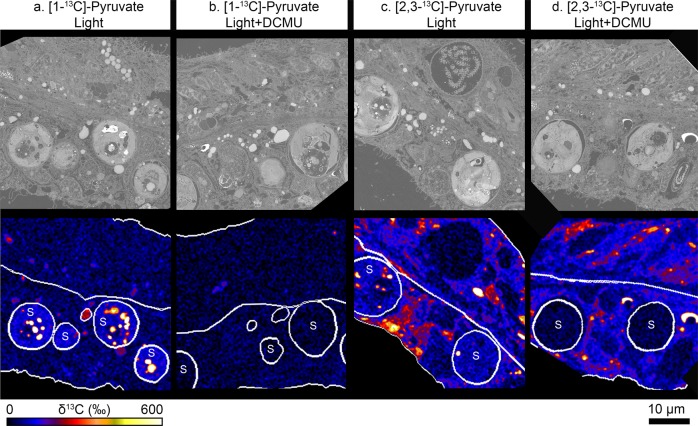
Fig. 1. Isolating host anabolism using position-specific 13C-labelling of pyruvate.
Corals were incubated with differentially labelled pyruvate, in the presence or absence of the photosynthetic inhibitor DCMU, for 12 h. Scanning electron microscopy (top row) images and their correlative NanoSIMS image (bottom row) are shown for corals labelled with a [1-13C]-pyruvate in the light; b [1-13C]-pyruvate in the light + DCMU; c [2,3-13C]-pyruvate in the light, and d [2,3-13C]-pyruvate in the light + DCMU. Circles labelled with ‘S’ show the position of the algal symbionts in their hosts’ oral gastrodermis, while unmarked circles show the position of host lipid bodies. Note that only a couple of host lipid bodies are circled for illustration purposes; these ROIs do not reflect the real abundance of lipids in the tissue, all of which were included in the analysis. See text for an explanation of the differential labelling patterns.

