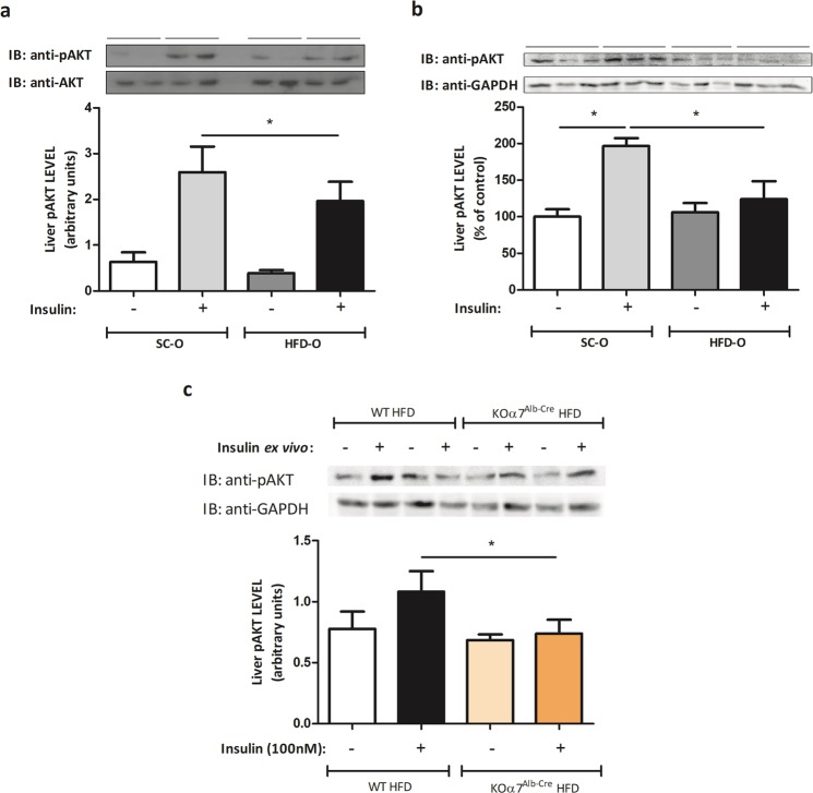
Figure 3.
Impairment of the insulin signalling in the obese mother’s offspring. Hepatic pAKT protein levels were evaluated by Western blot in control and obese dams’ offspring (P28) after a bolus injection of saline or regular insulin (5 UI) through the abdominal cava vein. The expression of the protein was measured after 45 seconds of insulin administration. The percent expression of control (AKT) is shown (means ± SD, n = 6 per group). (a) Hepatic pAKT protein levels were evaluated by Western blot in control and obese dams’ offspring (P28) after an ex vivo experiment where the tissue was treated with insulin (100 nM) for 10 minutes in the cell media. The percent expression of control (GAPDH) is shown (means ± SD, n = 6 pups for SC-O and n = 4 pups for HFD-O). (b) Hepatic pAKT protein levels were evaluated by Western blot in KOα7Alb-Cre and WT mice after an ex vivo experiment where the tissue was treated with insulin (100 nM) for 10 minutes in the cell media. The percent expression of control (GAPDH) is shown (means ± SD, n = 6 pups for SC-O and n = 4 pups for HFD-O). (c) Statistical significance was analysed by Student’s t-test for analysis of two groups (*p < 0.05).

