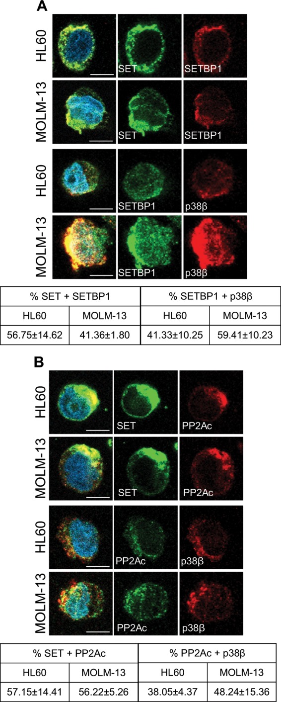
Fig. 3. p38β acts as a SET stabilizing protein.

a Immunofluorescence analysis of SET (green) and SETBP1 (red), and SETBP1 (green) and p38β (red), in HL60 and MOLM-13 cells. Nuclei were stained with DAPI. Quantification of colocalization fluorescence. (b) Immunofluorescence analysis of SET (green) and PP2Ac (red) and PP2Ac (green) and p38β (red), in HL60 and MOLM-13 cells. Nuclei were stained with DAPI. Quantification of colocalization fluorescence. Immunofluorescences were visualized by confocal microscopy. The results are expressed as mean values ± SEM. Experiments were performed in triplicate four times. *p < 0.05, **p < 0.01. Scale bar represents 5 µm.
