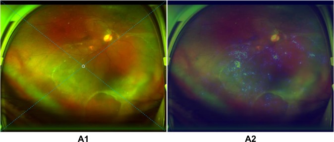
Fig. 7. Example of an ultra-widefield fundus image with a mistaken circle generated according to a heatmap.
A circle which should have been a downward arrow in image A1 of inferior retinal detachment is established mistakenly based on the highlighted region in heatmap A2. The arrow/circle is used to instruct patients in preoperative posturing to reduce the progression of retinal detachment between detection and treatment. The dotted diagonal line in the image is used to divide the retina into four quadrants (superior, inferior, left, and right).

