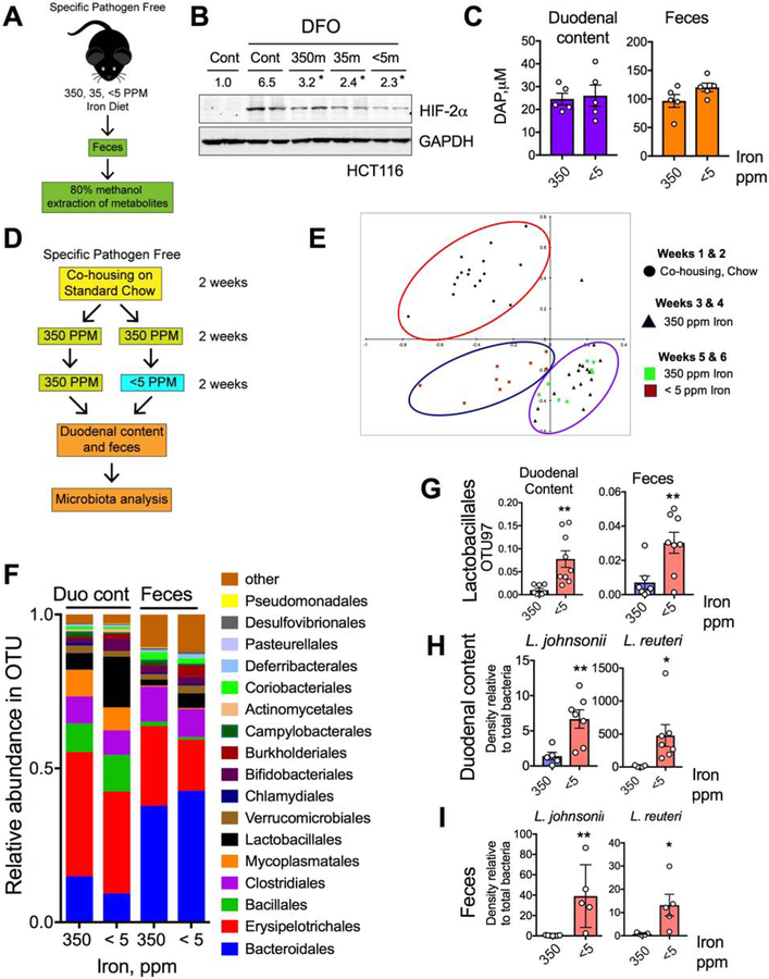
Figure 5. Low-iron diet potentiates HIF-2α inhibition by microbial metabolites.
A) Schematic of fecal metabolite extraction from SPF mice fed with 350-, 35- and < 5-ppm iron diet for 2 weeks and B) HIF-2α Western analysis in HCT116 cells treated with or without DFO and metabolites (350m, 35m or <5m respectively). C) DAP concentration in duodenum and feces from wild type fed with 350- and <5-ppm iron diet for two weeks. D) Schematic of the experimental set up for bacterial community analysis. E) Principal component analysis (PCA) of fecal bacterial population from 16S rRNA sequencing data in mice fed with 350-ppm and < 5-ppm iron diet. F) 16S rRNA sequencing bacterial community analysis from duodenal content and feces. G) Comparative analysis of genus Lactobacillales by OTU97 (97% sequence similarity) values in duodenal content and feces. Comparative analysis of L. johnsonii and L. reuteri density by species specific PCR from H) duodenal content and I) feces.
All data are mean ± SEM. t-test for the corresponding panels (C, G, H, I). Western analyses (B): Images were analyzed by Image J software from three independent experiments, representative image shown. Statistical significance compared with DFO-only treatment group. * P < 0.05, ** P < 0.01.

