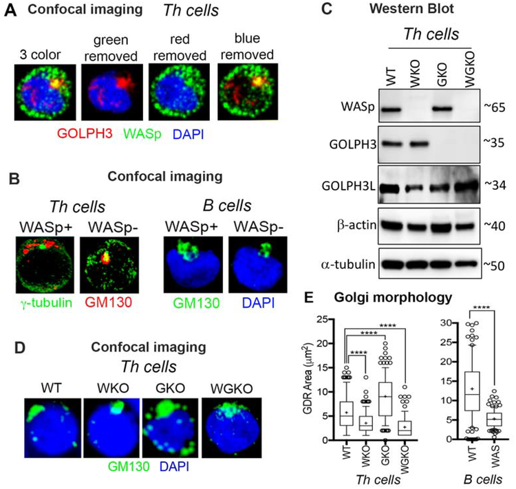
Figure 1. WASp is required to maintain the physiological Golgi morphology in T and B cells.
(A) Confocal immunofluorescence images of the human, primary Th cells triple-labelled with the indicated reagents. Displayed images are the collapsed composites of 20–30 z-stack images acquired at x63 magnification, and are representative of at least 50 cells for each condition. (B) confocal images of Th cells and B cells (WASp+, WASp−) dual-labelled with the indicated reagents showing Golgi morphology under basal, steady-state conditions. (C) Western blot showing the presence or absence of the indicated protein in 4 isogenic Th cell lines lacking either WASp (WKO), GOLPH3 (GKO), both WASp and GOLPH3 (WGKO), or wild-type (WT). (D) Confocal immunofluorescence images of the Golgi morphology in the same 4 Th cell lines shown in panel C and B cells (WT and WAS). (E) Box-and-whisker plots (whiskers@10–90%, horizontal bar denotes median, “+” denotes mean) generated from 3D (z-stack) images of at least n=100 cells from n=3 independent experiments. p**** ≤ 0.0001, Mann-Whitney nonparametric.

