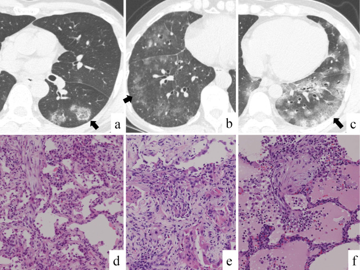
Figure 3.
The chest computed tomography findings and histologic findings of viral pneumonia. Patchy ground-glass opacities (GGOs) and nodules in a 44-year-old man with human parechovirus pneumonia (a). Bilateral GGOs and centrilobular nodules in a 72-year-old woman with mixed viral pneumonia due to respiratory syncytial virus, human parechovirus, and human parainfluenza virus (type 1) (b). Bilateral GGOs and consolidation in a 42-year-old man with mixed viral pneumonia due to influenza A virus, respiratory syncytial virus, and human parechovirus (c). Arrows indicate areas where tissue samples were obtained. Histological findings obtained via transbronchial lung biopsy (d, e) and video-assisted lung biopsy (f) showed organization and swollen pneumocytes. Histological findings obtained via surgical lung biopsy showed organization, swollen pneumocytes, and pulmonary edema (f). The photos of histological specimens in panels d, e, and f respectively correspond to the patients in panels a, b, and c.

