Fig. 1.

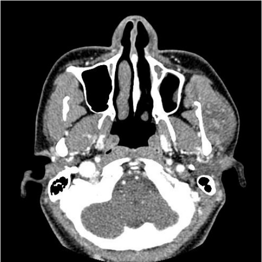
In the enhanced computed tomography image, a 2.8×2.8×1.8 cm heterogeneously enhancing mass in the left masseter muscle can be observed.

In the enhanced computed tomography image, a 2.8×2.8×1.8 cm heterogeneously enhancing mass in the left masseter muscle can be observed.