Fig. 3.

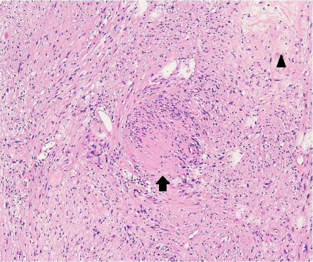
Photomicrograph (H&E, ×100) reveals that the tumor consists of focal cellular (Antoni A, black arrow) areas and hypocellular (Antoni B, black arrowhead) areas with vacuolar degeneration.

Photomicrograph (H&E, ×100) reveals that the tumor consists of focal cellular (Antoni A, black arrow) areas and hypocellular (Antoni B, black arrowhead) areas with vacuolar degeneration.