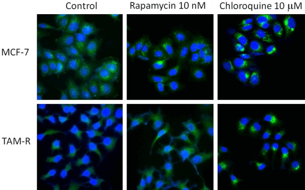
Figure 3.

Autophagy fluorescent microscopy of LC-3 in MCF-7 and TAM-R cells in the control, rapamycin, and chloroquine. MCF-7 and TAM-R cells grown on sterile glass cover slip in 6-well plates in IMEM-5%FBS were treated with rapamycin 10 nM and chloroquine 10 mM for 24 h and then stained with CytoID green dye for autophagosome and Hoechst 33342 for nuclei. The immunofluorescence staining was visualized with an Olympus (IX81) inverted fluorescent microscope. Imaging: FITC and DAPI (60 ×).
