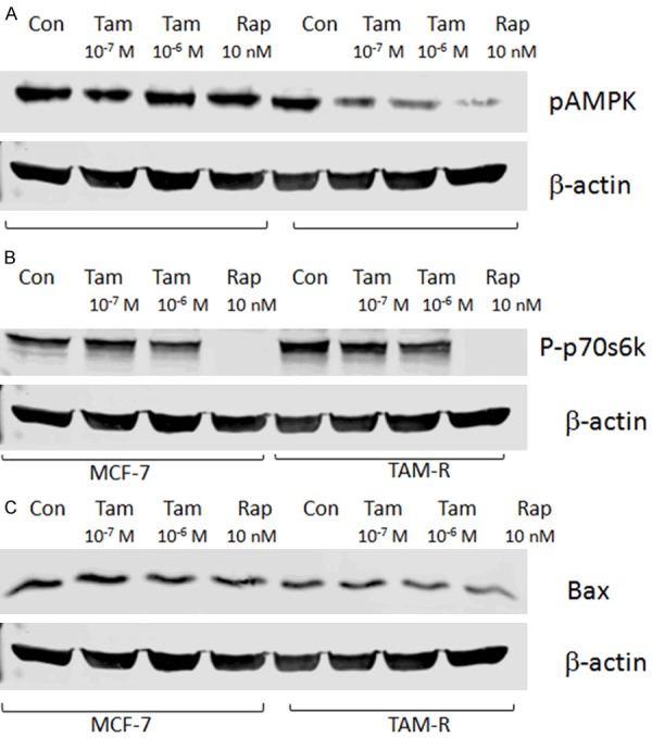
Figure 4.

Tamoxifen therapy altered the autophagy and apoptosis markers in the MCF-7 and TAM-R cells. The MCF-7 and TAM-R cells were plated in 60-mm dishes with 5% FBS IMEM and treated with tamoxifen (10-7 and 10-6 M), and rapamycin (10 nM) in a culture medium for 24 hours when the cells were about 80% of confluence before western blotting. Bar graphs indicate the relative levels of pAMPK, Bax, and P-p70s6k normalized to β-actin. A: The effects of tamoxifen on the expression of pAMPK in the MCF-7 and TAM-R cells; B: The effects of tamoxifen on the expression of P-p70s6k in MCF-7 and TAM-R cells; C: The effects of tamoxifen on the expression of BAX in the MCF-7 and TAM-R cells.
