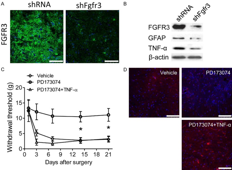
Figure 5.

Spinal TNF-α synthesis participates in FGFR3 induced mechanical hypersensitivity. (A) Intrathecal injection of SNI models with shFgfr3 or control lentivirus. Immunofluorescence images showed the expression of FGFR3 in the ipsilateral dorsal spinal cord at 14 days after surgery. (B) Expression levels of FGFR3, GFAP and TNF-α in the ipsilateral spinal dorsal horn were measured by western blot assays 14 days after shFgfr3 or control lentivirus intrathecal treatment. Β-ACTIN was used as control. (C) Intrathecal injection of SNI models with vehicle or PD173074 or PD173074 combined with TNF-α. Withdrawal thresholds were measured and compared between groups in the indicated days after surgery. Data represent the mean ± SEM. *P<0.05 vs. sham mice at the corresponding time point. n = 6/group. (D) Expression levels of GFAP in the ipsilateral spinal dorsal horn were measured by immunofluorescence in the rat treated as (C).
