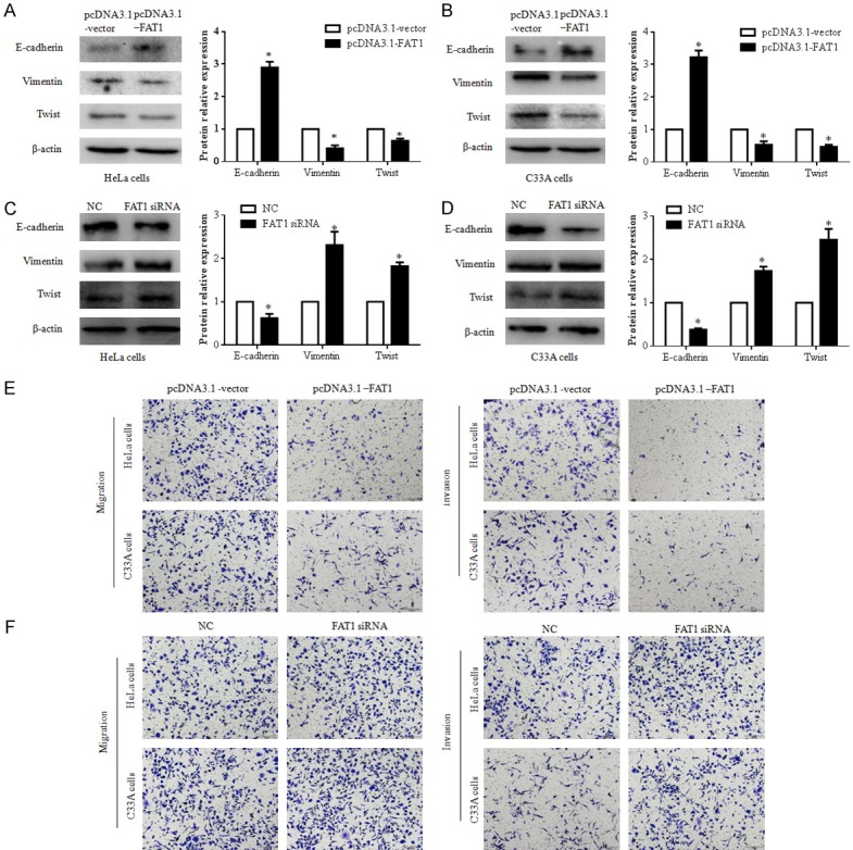
Figure 2.

FAT1 suppressed EMT, migration, and invasion of HeLa and C33A cells. (A and B) FAT1 supressed EMT of HeLa and C33A cells. The expression of E-cadherin, vimentin, and TWIST in HeLa (A) and C33A cells (B) were detected by western blot after these cells were treated with pcDNA3.1-FAT1 or pcDNA3.1-vector plasmid for 48 h, respectively. The histograms illustrate quantitative analysis of the E-cadherin, vimentin, and TWIST protein levels, which were normalized to the β-actin levels. *P < 0.05 vs. the pcDNA3.1-vector group. (C and D) Silenced FAT1 promotes the EMT of HeLa and C33A cells. The expression of E-cadherin, vimentin, and TWIST in HeLa (A) and C33A cells (B) were detected by western blot after these cells were treated with NC or FAT1 siRNA for 48 h, respectively. The histograms illustrate the quantitative analysis of the E-cadherin, vimentin, and TWIST protein levels, which were normalized to β-actin levels. *P < 0.05 vs. the NC group. (E and F) The effect of FAT1 on HeLa and C33A cell migration (E) and invasion (F) capacity was analyzed using a transwell assay. These cells were transfected with the pcDNA3.1-FAT1/pcDNA3.1-vector plasmid or NC/FAT1 siRNA for 24 h, respectively.
