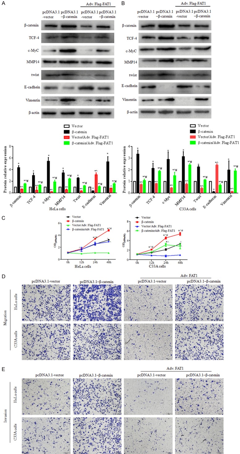
Figure 4.

Upregulation of β-catenin partly reversed FAT1’s effect on proliferation, migration and invasion. HeLa and C33A cells were transfected with the vector and β-catenin plasmid for 12 h, then infected with adv. Flag-FAT1. (A and B) Western blotting showed the expression levels of β-catenin, TCF-4, c-Myc, MMP14, TWIST, E-cadherin, and vimentin in HeLa and C33A cells. The histograms illustrate the quantitative analysis of the pβ-catenin, β-catenin, TCF-4, c-Myc and MMP14 protein levels, which were normalized to the β-actin levels. *P < 0.05, vs. the pcDNA3.1-vector group. ^P < 0.05, vs. the β-catenin group. #P < 0.05, vs. the Vector/Adv.Flag-FAT1 group. (C) CCK-8 assays were used to analyze HeLa (left panel) and C33A (right panel) cell viability. *P < 0.05 vs. the vector group. ^P < 0.05 vs. the vector/adv. Flag-FAT1 group. #P < 0.05 vs. the β-catenin/adv. Flag-FAT1 group. (D and E) A transwell assay was used to evaluate HeLa and C33A cell migration (D) and invasion (E) capacity with co-expression of β-catenin and FAT1. Treated cells were seeded in upper chamber for 24 h, respectively.
