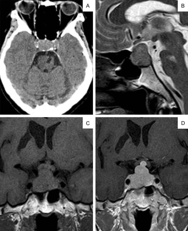
Figure 1.

Axial CT image shows the localized pituitary lesion that eroded the bone and invaded the cavernous sinus (A). The mass extended to the suprasellar and showed up as isointense on the T2-weighted sagittal MRI image (B). Pre-(C) and post-(D) contrast images showed the mass oppressing the optic chiasm. The mass was homogeneously enhanced with Gd-DTPA.
