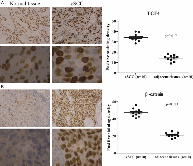
Figure 1.

The expression of TCF4 in cSCC tissues. Immunohistochemical analysis of TCF4 (A) and β-catenin (B) in cSCC cancer tissues.TCF4 and β-catenin positive cell nuclei are also detected in the cSCC (20 ×). Magnified images (100 ×) are shown at the bottom. Data is presented as the mean ± standard error of the mean of five picked individual fields.
