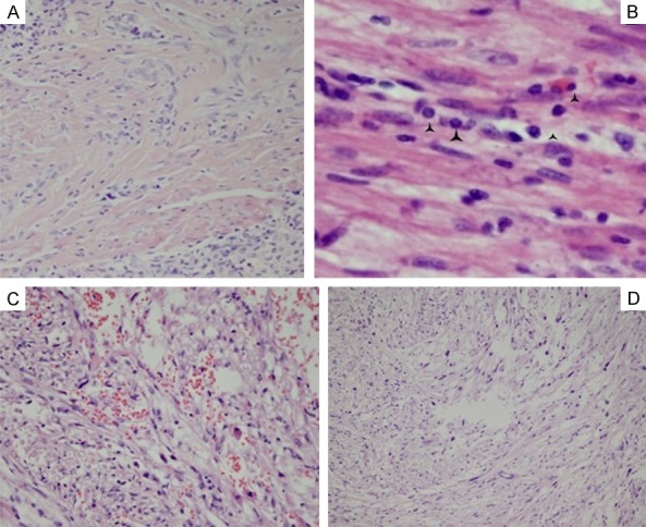
Figure 3.

The changes of interstitial stroma: (A) The hyaline change of stroma. The black angles in (B) showed the inflammatory cell infiltrate; (C) Scattered hemorrhage and edema; (D) Focal polymorphism with multinuclear giant cells. (A, D): 4 × 10; (C): 10 × 10; (B): 40 × 10.
