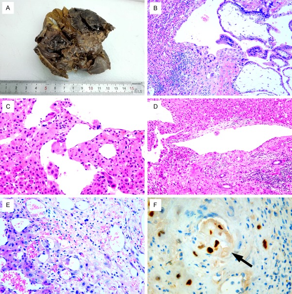
Figure 2.
A. Gross pathologic examination found a 5×5×4 cm reddish mass detected within the hepatic tissues on the cut surface. B. Histologically, the chorionic villi with trophoblasts eroded into the hepatic tissues (×100). C-E. A few trophoblasts can be detected in some hepatic veins (×200). F. HCG immnostaining showed positive reactivity in the trophoblasts in some small veins (×400, black arrow).

