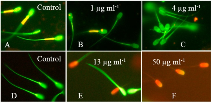
Figure 6.
Epifluorescence micrographs of boar sperm exposed to crude extract from A. calidoustus strain MH34. The sperm cells in the upper row were stained with the potentiometric dye JC-1, which stains membranes with high membrane potential ΔΨ orange and membranes with low ΔΨ green. Sperm cells in the lower row were stained with the viability stain calcein-AM + propidium iodide. Cells with intact plasma membranes are stained green, and cells with depleted plasma membrane integrity are stained red. Panel (A) shows sperm cells exposed to the solvent only exhibiting high ΔΨ in the mitochondrial sheath located in the midpiece of the sperm cells. Panels (B,C) show the concentrations for which 50% and 100% of sperm cells, respectively, exhibited depolarized mitochondria, as indicated by a decrease in orange emission from the mitochondrial sheath. Panel (D) shows control-exposed sperm cells with intact plasma membranes emitting green fluorescence. Panels (E,F) show sperm cells exposed to concentrations causing 50% and 100% depletion of the plasma membrane integrity, respectively.

