Figure 3.
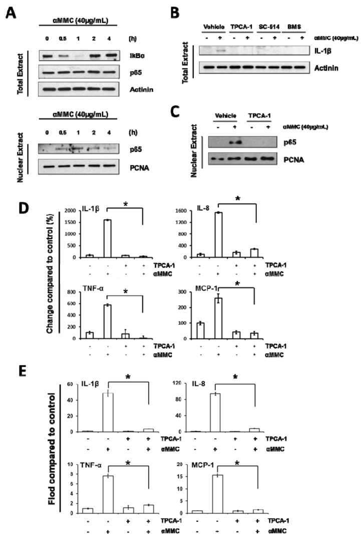
NF-κB activation is essential in α-MMC-induced inflammatory responses in THP-1 cells. (A) THP-1 cells were incubated with α-MMC (40 μg/mL) for various time intervals (0, 0.5, 1, 2, 4 h) and levels of IκBα and p65 proteins in the whole cell lysates and p65 protein in the nucleus were assayed by Western blot. Actinin and PCNA were employed as loading control for whole cell and nuclear extracts, respectively. (B) THP-1 cells were pretreated with specific IKKβ inhibitors TPCA-1 (5 µM), SC-514 (50 µM), BMS-345541 (2.5 µM) for 30 min, and then the cells were further treated with vehicle control or α-MMC (40 μg/mL) for 24 h. Western blotting was used to analyze the protein expression levels of IL-1β in whole-cell extracts. Actinin was employed as loading control. (C) THP-1 cells were exposed to vehicle or α-MMC (40 μg/mL) for 24 h after pretreatments with or without 5 μM TPCA-1 for 30 min. Western blot analysis of nuclei p65 is shown. PCNA was employed as loading control of nuclear extracts. (D) THP-1 cell cultures were pretreated with specific IKKβ inhibitor TPCA-1 (5 μM) for 30 min, follow by the addition of α-MMC (40 μg/mL) and further incubated for 24 h. The secretion levels of indicated cytokines in the cell culture supernatant were examined using corresponding ELISA kits. The results are expressed as the change in percentage compared to vehicle control group. The data represent the mean ± SD of three replicates. Significant differences: * p < 0.01. (E) THP-1 cells were incubated with specific IKKβ inhibitor 5 μM TPCA-1 for 30 min prior to the treatment of 24 h with α-MMC (40 μg/mL). The mRNA levels of the four pro-inflammatory cytokines were analyzed by RT-PCR. Results were expressed as fold-change compared to the vehicle control group. The data represent the mean ± SD of three replicates. Significant differences: * p < 0.01.

