Figure 4.
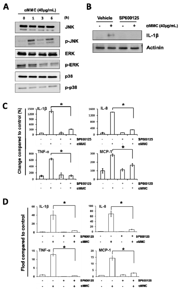
JNK activation is essential in α-MMC-induced inflammatory responses in THP-1 cells. (A) Western blot of THP-1 cells incubated with α-MMC (40 μg/mL) for different time intervals (0, 1, 3, 6 h). Whole-cell extracts were extracted and phosphorylated JNK, ERK, and p38 levels were analyzed by Western blot analysis using corresponding antibodies. (B) THP-1 cells were firstly treated with 15 μM JNK inhibitor SP600125 for 30 min, followed by adding of α-MMC (40 μg/mL) and further incubated for 24 h. Whole cell lysates were extracted and protein expression levels of IL-1β were analyzed by Western blot. Actinin was employed as an equal loading control. (C) THP-1 cells were exposed to vehicle or α-MMC (40 μg/mL) for 24 h after pretreatments with or without 15 μM JNK inhibitor SP600125 for 30 min. Secretions of IL-1β, IL-8, TNF-α, and MCP-1 in THP-1 cells culturing medium were detected by ELISA. The results are shown as a percentage of change compared to control group. Data represent the mean ± SD of three replicates. Significant differences: * p < 0.05. (D) THP-1 cells were incubated with 15 μM SP600125 for 30 min prior to the treatment of 24 h α-MMC (40 μg/mL). The mRNA levels of the indicated cytokines were analyzed by RT-PCR. Results are shown as fold-change compared to the control. Data represent the mean ± SD of three replicates. Significant differences: * p < 0.01.

