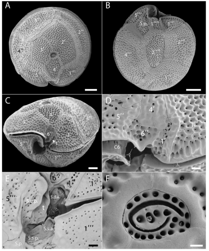
Figure 3.
Scanning electron microscopy (SEM) micrographs of G. belizeanus (strain CUB5G4). (A): Epitheca. (B): Hypotheca. (C): Ventral view. (D): Detail of the small ventral plates 1′ and 6′′. (E): Detail of sulcal plates. (F): Apical pore plate (Po) with the fishhook-shaped pore. Scale bars: A, B, C: 10 µm, D, E: 2 µm and F: 1 µm.

