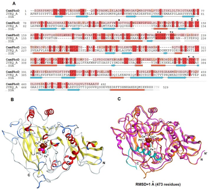
Figure 8.
(A) Modeling of the CamPhoD 3D structure. An alignment of the amino acid sequences of the alkaline phosphatase/phosphodiesterase CamPhoD from the marine bacterium C. amphilecti KMM 296 (GenBank ID: WP_043333989) and alkaline phosphatase (phosphodiesterase) D from B. subtilis (PDB ID: 2YEQ). The amino acid sequences identity and similarity (color boxed) and the secondary structure of the template are highlighted. Note: α-helixes = red sticks; β-structure = blue arrows; the binding of amino acid (aa) residues (Ca2+/Co2+) = *; the binding of conserved aa residues (Fe3+) = o; and the residue Cys 124 of the template = •. (B) The 3D structure model of CamPhoD with the reaction product Pi and metal ions in the active center (the protein structure is a ribbon diagram, Pi is in stick form, and Ca2+ is shown as spheres). (C) The 3D superimposition of the CamPhoD model (orange) and the template (PDB ID: 2YEQ) (shown in pink, with the blue C-terminal part).

