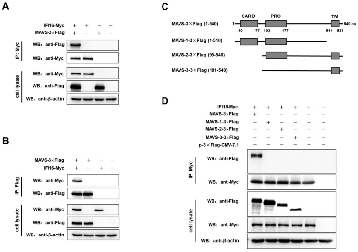
Figure 6.
IFI16 interacts with MAVS. (A,B) HEK-293T cells were co-transfected with IFI16-Myc and MAVS-3×Flag for 48 h. Cells were lysed, and the lysates were subjected to immunoprecipitation and analyzed by western blotting using indicated antibodies. (C) Scheme of MAVS protein and its mutants. (D) HEK-293T cells were co-transfected with IFI16-Myc and MAVS-3×Flag, MAVS-1-3×Flag, MAVS-2-3×Flag, MAVS-3-3×Flag, p3×Flag-CMV-7.1, and 48 h later, the cells were lysed and subjected to immunoprecipitation and analyzed by western blotting.

