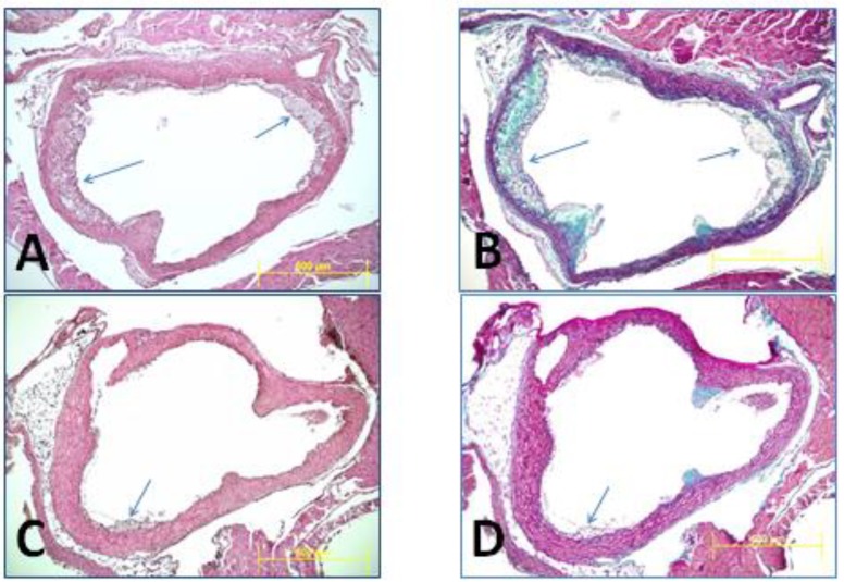
Figure 3.
Representative photomicrographs were taken at the beginning of aorta from one control mouse (A,B) and one wild rice fed mouse (C,D) illustrating atherosclerotic lesions (arrows). As it is seen in (A,B), atherosclerotic lesions are large and well established in the control mouse (arrows), while such advanced lesions are missing in the wild rice fed mouse (C,D). H&E staining (A,C); trichrome staining (B,D).

