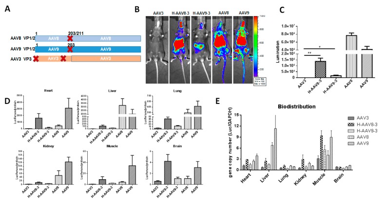
Figure 6.
Transduction of haploid vectors with VP3 from AAV3 and VP1/VP2 from AAV8 or AAV9. (A) The composition of AAV capsid subunits. Haploid AAV viruses were produced by co-transfection of two plasmids (one encoding VP1 and VP2 from AAV8, or AAV9, VP3 from AAV3). “X” represents start codon mutation. (B) Luciferase expression in the representative mice. 1 × 10¹⁰ particles of AAV vector were systemically administered into the mice. Imaging was performed at day 7. (C) The quantitation of liver transduction. The luciferase signal was measured and calculated. (D) Ex vivo luciferase expression of the tissues. The harvested tissues were lysed and analyzed by luciferase assay. (E) Bio-distribution of haploid vectors. The AAV genomic copy number was measured by qPCR assay. Each group contains five mice. Asterisks indicate a significant difference between the groups at the levels of * p < 0.05 and ** p < 0.01.

