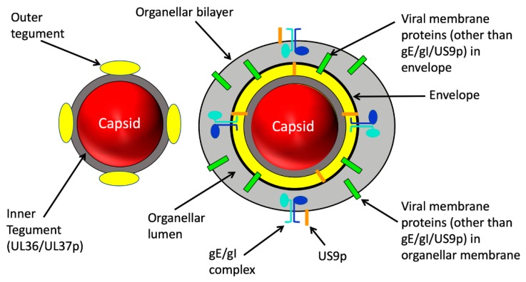
Figure 2.
Structure of alphaherpesvirus particles that utilize MTs during assembly and traffic. Left: The naked capsid. After assembly and packaging in the nucleus the capsid (red sphere) is delivered to the cytoplasm. The inner tegument proteins UL36p and UL37p (dark grey layer) provides the foundation for attachment of outer tegument subunits (yellow) prior to or during cytoplasmic envelopment (not shown). Right: the organelle-enclosed enveloped virion (OEV). Budding of the naked capsid into the lumen (light grey space) of a cytoplasmic organelle is accompanied by completion of the outer tegument (yellow) and generates a mature infectious enveloped virion within the organellar lumen. Multiple virally encoded membrane proteins (green bars) reside in the viral envelope and surrounding organellar membrane. For the purposes of this review three virally encoded membrane proteins are shown in detail: the gE/gI glycoprotein heterodimer (subunits shown in light blue and dark blue) and the US9p protein (orange bar). Note that US9p has essentially no lumenal domain. US9p and the gE/gI complex in the organellar bounding membrane project tails into the cell cytoplasm.

