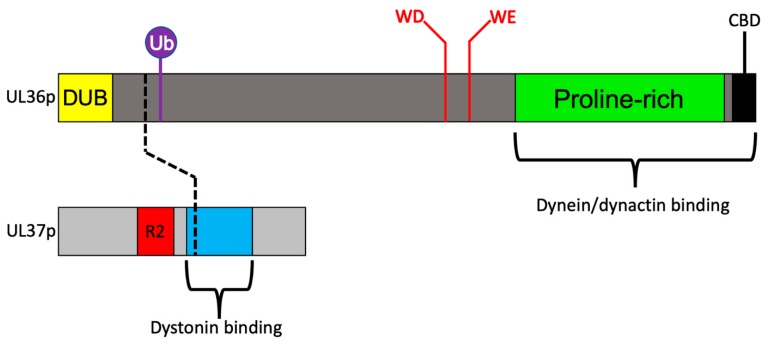
Figure 3.
The UL36p and UL37p inner tegument proteins. Upper bar: The UL36p polypeptide. Regions discussed in this review are annotated as follows: DUB (Deubiquitinase) domain: yellow. Ub (Ubiquitin) attachment site: purple lollipop. WD/WE (tryptophan-acidic) motifs: red text. Proline-rich region: green. CBD (carboxy terminal capsid binding domain: black (additional capsid binding domains exist in UL36p but are not shown). Region sufficient for dynein/dynactin interaction is bracketed. Lower bar: UL37p polypeptide. R2 surface region: red. Site of dystonin binding: blue. Broken black line connects regions of UL36p and UL37p implicated in their association.

