Figure 1.
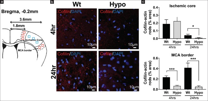
Cofilin-actin rod formation was reduced by therapeutic hypothermia. (a) Schematic showing where regions of interests were sampled as they represented the ischemic core (3.6 mm from Bregma, blue squares) and the brain margins supplied by the middle cerebral artery (MCA border 1.8 mm from Bregma, red squares). (b) Representative images of cofilin (red) and DAPI (blue) staining taken from the MCA border at 4 and 24 h postmiddle cerebral artery occlusion among normothermic and hypothermic animals. Fewer rods were observed in the hypothermia brain. (c) Quantification of the brain regions shows the percentage area occupied by cofilin-actin rods within the ischemic core and middle cerebral artery (MCA) border at 4 and 24 h. Within the core, rods increased in both groups at 4 h and markedly reduced at 24 h, with significant reduction in the hypothermia group at 24 h. Within the MCA border, rods were more marked at 24 h compared to 4 h, but at both time points, rods were reduced in the hypothermia group (*P < 0.05, ***P < 0.01)

