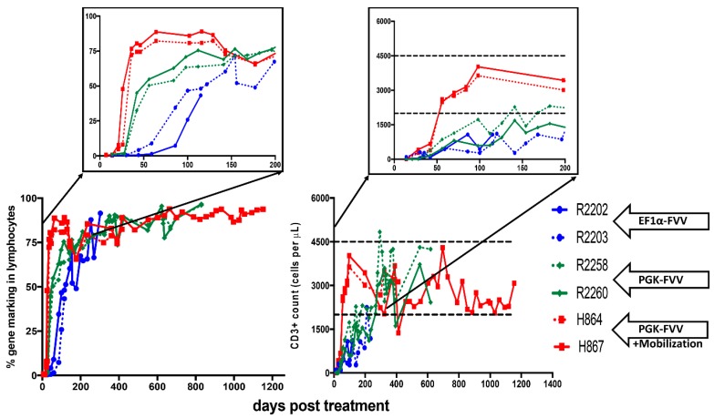
Figure 1.
Immune reconstitution in foamy virus vector (FVV) treated X-linked severe combined immunodeficiency (SCID-X1) dogs: Bottom left graph shows % gene corrected lymphocytes in peripheral blood of various animals. Top left inset highlights the early kinetics of gene marking in treated animals (blue vs. green vs. red lines). The mobilized dog H867 had a stable level of gene marking almost 1260 days post treatment. Bottom right graph represents absolute number of CD3+ T lymphocytes in peripheral blood of treated dogs. Top right inset emphasizes the days required to attain normal numbers of absolute T lymphocytes (blue vs. green vs. red lines; dashed black lines shows counts in healthy dogs). H867 maintained normal levels of CD3+ T cell counts for over three years. Data in this figure was reproduced from previous studies [7,8] and contains extended data on H864 and H867. R2202 and R2203 were part of the cohort of five dogs from our first study [7] and R2258, R2260, H864 and H867 were part of our second study [8], additional details are included in the text. EF1α-FVV: elongation factor 1 α promoter (EF1α-GFP-2A-γC) carrying foamy virus vector (FVV); PGK-FVV: human phosphoglycerokinase (PGK-mCherry-γC) promoter carrying FVV.

