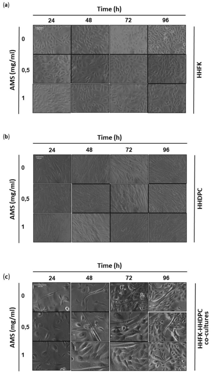
Figure 2.
Cytomorphological analysis on cell monolayers. Representative microphotographs by phase-contrast light microscopy at a 100× magnification (10× objective and a 10× eyepiece) of HHFK (a) and HHDPC (b), and at a 200× magnification (20× objective and a 10× eyepiece) of HHFK-HHDPC co-cultures (c) treated for the indicated times (from 24 to 96 h) with 0.5 and 1 mg/mL of AMS nutraceutical, as indicated. The shown images are representative of three independent experiments.

