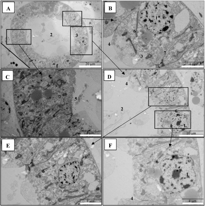
Figure 4.
Digestive tract in control brine shrimp larvae appeared normal, with intact cellular membranes. Transmission electron microscopy (TEM) analysis of control group brine shrimp (A. franciscana) larvae after 48 h. (A–F): The digestive tract epithelial enterocytes appeared normal with an intact mitochondrion, nucleus, rough endoplasmic reticulum (RER), and intercellular junctions. Legend: (1) nucleus; (2) midgut lumen; (3) midgut enterocytes; (4) tight junction; (5) microvilli; (6) mitochondria; (7) rough endoplasmic reticulum (RER).

