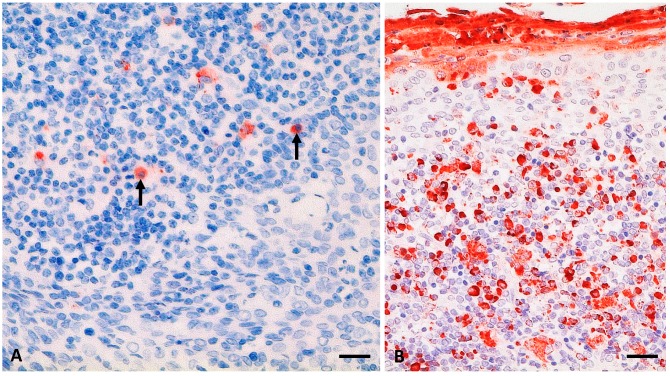
Figure 2.
PPRV antigen (-ag) detection (in red) in palatine tonsil from an alpaca (A2) at 28 days post infection (dpi) (A; see also Table 2) and a goat (positive control; G1) at 9 dpi (B) after experimental infection with the PPRV Kurdistan/2011 strain. (A) mild PPRV-ag detection (6% to 25% positive cells) in single lymphoid cells of an enlarged section of a lymphoid follicle of the tonsil from A2, including lymphocytes (arrows); (B) positive control, palatine tonsil section from a clinically severely affected goat with severe accumulation of PPRV-ag (>75% positive cells) in mononuclear lymphoid cells as well as epithelia and desquamated material. Immunohistochemistry, monoclonal mouse anti-PPRV-Np (IDvet); scale bar indicates 20 µm.

