Fig. 2.

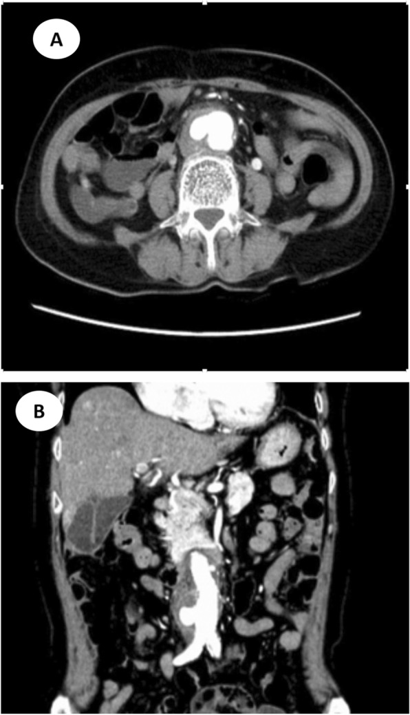
Computed tomographic angiography (CTA) of abdomen before surgery (a) Axial view (b) Coronal view. CTA during contrast phase showed abdominal aortic aneurysm with atherosclerotic changes, together with mural thrombus and penetrating ulcer

Computed tomographic angiography (CTA) of abdomen before surgery (a) Axial view (b) Coronal view. CTA during contrast phase showed abdominal aortic aneurysm with atherosclerotic changes, together with mural thrombus and penetrating ulcer