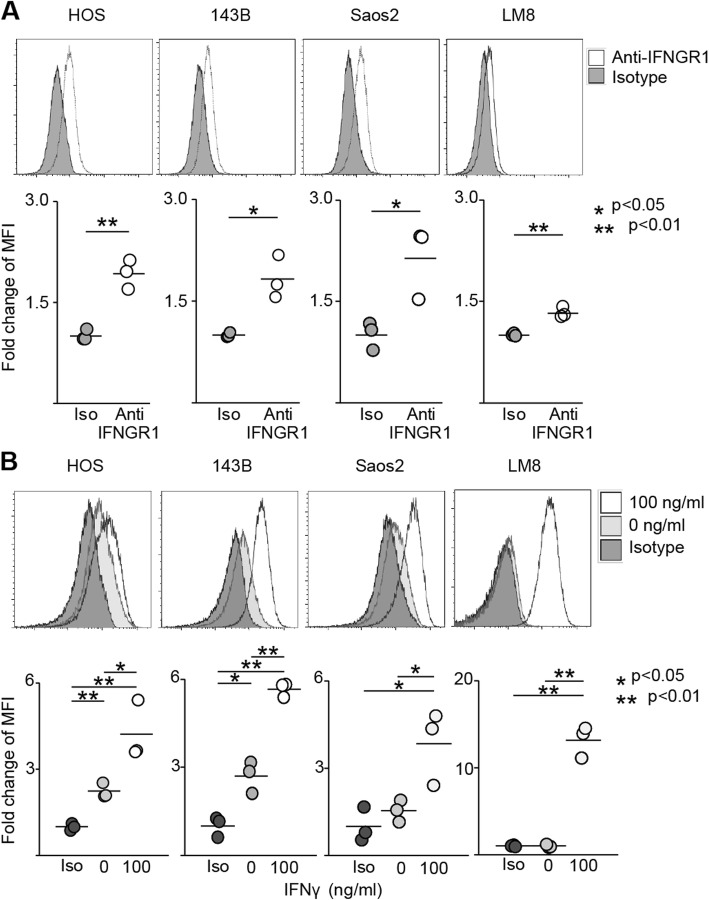
Fig. 1.
IFNγ increases PD-L1 expression in osteosarcoma cell lines. Surface markers of human and murine osteosarcoma cell line were evaluated by flow cytometry. The upper row shows representative specimen. In the lower row, each specimen is plotted (n = 3), and the average value is indicated by a horizontal bar. a Expression of IFNGR1 in each cell line. b Expression of PD-L1 in each cell line. Iso-type control, anti-PD-L1 staining with/without IFNγ were evaluated

