Fig. 2.
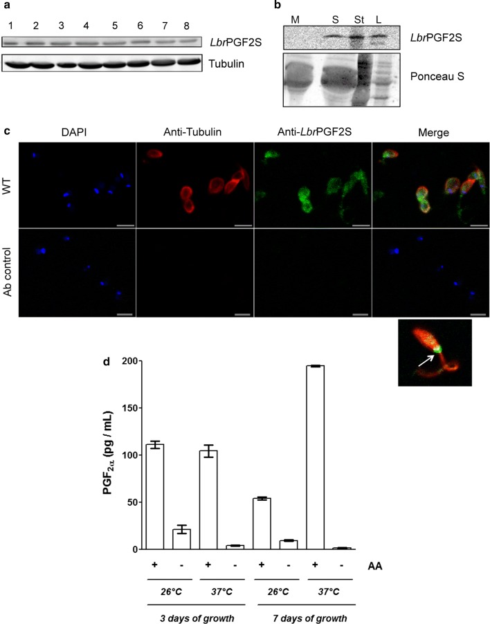
LbrPGF2S expression and secretion in L. braziliensis promastigotes. a Evaluation of LbrPGF2S protein levels in L. braziliensis promastigotes grown in axenic culture for 8 days using polyclonal anti-LbrPGF2S. Anti-Tubulin antibody was used for protein loading control. b Secretion of LbrPGF2S by promastigotes in the axenic medium. Immunoblots were used to detect LbrPGF2S in logarithmic (L) and stationary (St) phase promastigotes, 3 and 7 days of culture, respectively, or in the supernatant of the stationary phase culture (S) using anti-LbrPGF2S. Parasite-free M199 medium (M) was used as a negative control. Blots were stained with Ponceau S (lower panel) for protein loading control. c Immunofluorescence imaging used to detect LbrPGF2S in wild type promastigotes (at stationary phase). LbrPGF2S (CF488A, green); tubulin (Alexa555, red); DNA was stained with DAPI (blue). An image of a single promastigote is shown bottom right; a strong fluorescence signal appears at the flagellar pocket (white arrow). Ab control: parasites submitted to the same labelling protocol without primary antibodies. d Dosage of prostaglandin F2a synthesized by promastigotes in the presence (+) or absence (−) of 66 µM arachidonic acid (AA), measured by EIA assay. Quantification was performed with promastigotes at day 3 (logarithmic phase) or 7 (stationary phase), at 26 °C or 37 °C, as indicated below the x-axis. Data are means ± standard deviation from three replicates. Scale-bars: c, 5 µm

