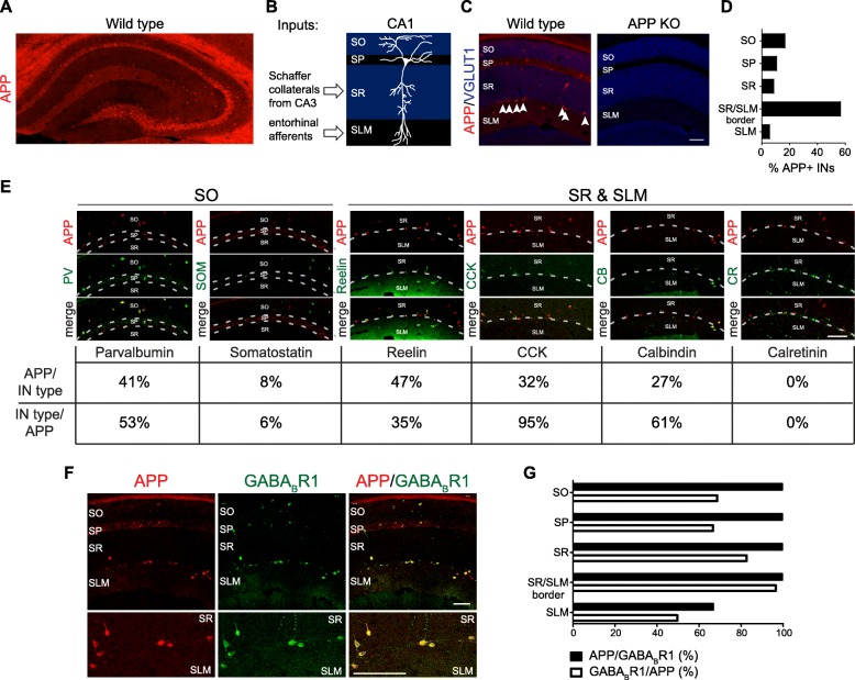
Fig. 1.
APP expression in interneurons of CA1 hippocampus. a Representative confocal image of whole hippocampus from 5-week-old wild type mouse section immunostained for APP. b Schematic of the CA1 subfield of the hippocampus. c Representative confocal images of hippocampal CA1 subfield of 5-week-old wild type or App KO mouse hippocampal sections immunostained for APP and excitatory presynaptic marker VGLUT1. Arrow heads denote APP-positive interneurons at SR/SLM border. d Quantification of the laminar distribution of a total of 54 APP-positive interneurons in CA1 examined over 4 sections from 4 different mice. e Representative confocal images of 5-week-old wild type mouse hippocampal sections co-stained with APP and interneuron markers (top panels) and quantification of their overlap (bottom panels). For each marker, a total of at least 90 APP-positive interneurons from at least 6 total sections from 2 different mice were examined. f Representative confocal images 5-week-old wild type mouse hippocampal sections co-stained with APP and GABABR1. The GABABR1 antibody does not distinguish 1a vs 1b; whereas only 1a is an APP binding partner. g Quantification of the overlap between APP-positive and GABABR1-positive GABAergic cells in CA1 laminae. A total of 54 APP-positive cells and 64 GABABR1-positive were examined over 4 sections from 4 different mice. IN = interneuron; SO = stratum oriens; SP = stratum pyramidale; SR = stratum radiatum; SLM = stratum lacunosum-moleculare. Scale bars = 100 μm

