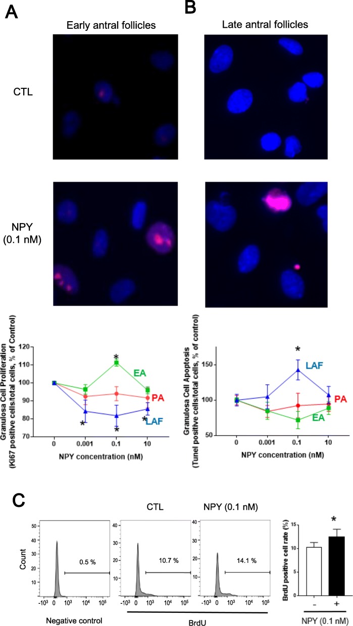
Fig. 4.
NPY induces granulosa cell proliferation in EA and reduced that in LAF. Granulosa cells of PA, EA and LAF were isolated separately using rats primed with eCG, as described in the methods section. a, b Granulosa cells were incubated with various concentration of NPY for 24 h and immunofluorescence of Ki67 (proliferation) and TUNEL assay (apoptosis) were performed. NPY induced Ki67-positivity of granulosa cells in EA (0.1 nM), while reduced that in LAF (0.001, 0.1, 10 nM; A). NPY induced TUNEL-positivity of granulosa cells in LAF (0.1 nM; B). c Granulosa cells from EA were incubated with NPY (0.1 nM) for 24 h and BrdU (10 μM) for last 4 h, and BrdU positive cells were detected by flow cytometry. Numbers displayed indicate percentage of single BrdU positive cells. NPY (0.1 nM) induced BrdU-positive granulosa cells in EA. These results suggest that NPY regulates granulosa cell proliferation and apoptosis in a follicular stage-dependent manner, inducing granulosa cell proliferation in early antral stage but apoptosis in late antral stage. Results are expressed as mean ± SEM of independent replicates (A, n = 5; B, n = 4; C, n = 4) and analyzed by two-way ANOVA and Tukey’s multiple comparison a,b and paired t-test c. *, p < 0.05 (vs. without NPY). Representative pictures and flow cytometry results are shown

