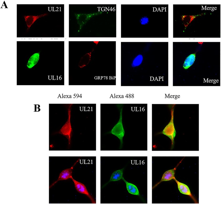
Fig. 6.
Colocalization of pUL16 and pUL21 in HEK 293 T cells. A: Localization of pUL21 (red) and pUL16 (green) alone with TGN46 (green) and GRP78 BiP (red) as cell markers in HEK 293 T cells. Images were captured under fluorescence microscopy using a 40× objective. B: Colocalization of pUL16 and pUL21 in HEK 293 T cells with pCAGGS-UL21-Flag (red) and pCMV-Myc-UL16 (green). Images were captured under fluorescence microscopy using a 40× objective

