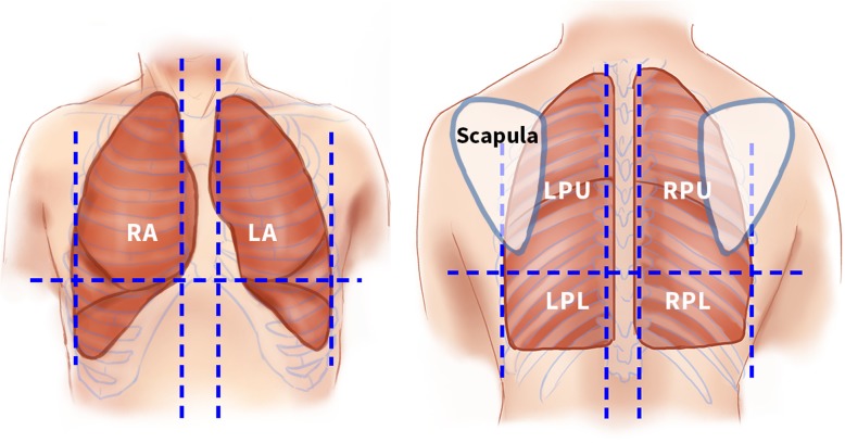
Fig. 2.
Anatomical zones scanned in lung POCUS. Illustrations of the front (left) and back (right) of the chest showing the six anatomical zones scanned. LA left anterior, LPL left posterior lower, LPU left posterior upper, POCUS point-of-care ultrasound, RA right anterior, RPL right posterior lower, RPU right posterior upper

