Figure 1.

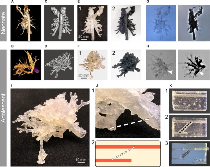
Patient‐specific in vitro pulmonary artery atresia model. Neonatal and adult vasculature with pulmonary artery atresia pathology were imaged via computed tomography (CT) (A) and 3‐dimensional (3D) rotational angiography (B). The vessel lumens were converted into 3D stereolithography files with 1‐mm thickness for neonatal and adult patients (C and D, respectively). These digital models were subsequently 3D printed (E and F) using different synthetic resins, including Clear Resin (1) and Flexible Resin (2) to pinpoint the most suitable material for the phantom. Validation using x‐ray angiography using iodine‐based contrast agent was performed on each patient model (G and H) without (left) and after (right) contrast addition. Arrows in (H) mark the vascular atresia. A simplified phantom was derived from the adult patient model by isolating the 3D area that included the occluded vessel and a functional vessel (I). Zoomed view of this area (J, 1) and the extrapolated stenotic phantom (J, 2) are shown. Proof‐of‐concept synthetic models of the phantom were created to evaluate its use (K). Untreated model (1), anastomosed vessels model (2), and stented model (3), mimicking the proposed treatment procedure, were 3D printed using Clear Resin for phantom optimization and iteration purposes.
