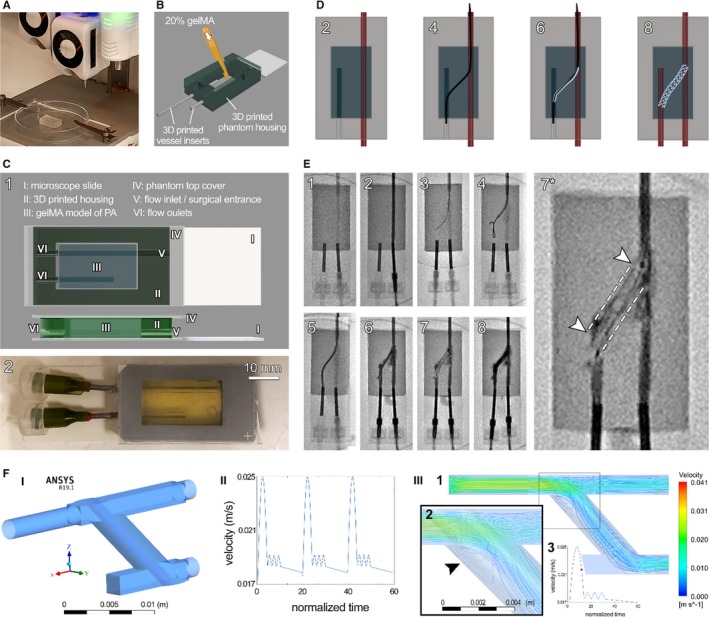
Figure 2.

Bioprinted phantom for recanalization of atretic vessels. A commercial 3‐dimensional (3D) bioprinter (CELLINK Bio X) was used to generate the biological model of tetralogy of Fallot with major aortopulmonary collateral arteries (MAPCAs) (A), which were then assembled into the 3D‐printed housings. A second approach, gelatin methacrylate (gelMA) casting directly into a 3D‐printed housing, was used to generate a second batch of phantoms (B). Render of the assembled phantom (C) shows its key parts (I‐VI, C‐1), with a fully assembled example shown below (C‐2). Schematic of the proposed recanalization procedure (D), showing flow in only the open vessel (2), catheter bridge (4), stent introduction (6), and stent deployment with flow restored in both vessels (8). Catheterization laboratory capture of the full procedure (E) shows the major (1–8) steps, starting from device setup and flow test (1–2) through establishment of bridge between the vessels (3–5), stent introduction (6) and deployment (7), and finally restored flow into both vessels (8). Zoom‐in of the deployed stent before flow reestablishment in both vessels is shown in (7*). The procedure was repeated 3 times. F, Computational fluid dynamic modeling of the stented atretic model, demonstrating (I) the computer‐aided design (CAD) model, (II) normalized flow velocity waveform over the cardiac cycle prescribed, and (III, 1–3) the characteristic flow pattern during the downstroke of systole. Arrow in III‐2 designates a turbulent flow region at the entry of the recanalized connection. III‐3 shows one pulse of velocity waveform and the red circle highlights the time point at which the flow data were acquired. PA indicates pulmonary artery.
