Figure 1.

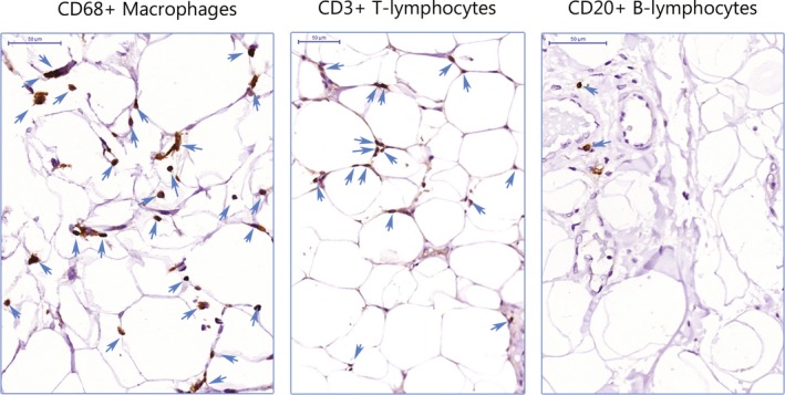
CD68+ macrophages, CD3+ T lymphocytes, and CD20+ B lymphocytes. Immune cells stained in brown and marked with a blue arrow infiltrated into the perivascular adipose tissue (bar=50 µm).

CD68+ macrophages, CD3+ T lymphocytes, and CD20+ B lymphocytes. Immune cells stained in brown and marked with a blue arrow infiltrated into the perivascular adipose tissue (bar=50 µm).