Figure 1. .
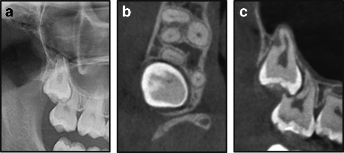
An impacted right maxillary third molar shown in the panoramic image (a); CBCT image in the axial plane (b); and CBCT image in the sagittal plane (c). The CBCT shows severe ERR with ERR of the entire distobuccal root of the second molar. CBCT, cone beam CT; ERR, external root resorption.

