Figure 2. .
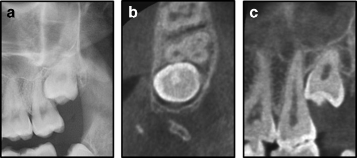
An impacted left maxillary third molar shown in the panoramic image (a); CBCT image in the axial plane (b); and CBCT image in the sagittal plane (c). The CBCT shows close relation between the crown of the third molar and the root of the second molar, but no ERR. CBCT, cone beam CT; ERR, external root resorption.

