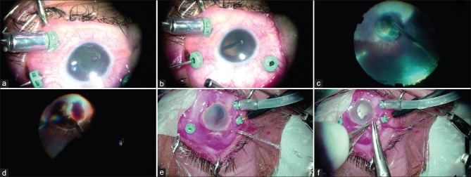
Figure 1.
(a) Sclerostomy ports are made at 3 mm from the limbus following which the cutter is used to engage the zonules and truncate it; this also in parallel creates dehiscence in the posterior capsule. The cutter is also used to mechanically dislocate the nucleus while cutting the zonules; this allows convenient visualization of the site of interest. (b) Once the zonulectomy is completed, the nucleus spontaneous dislodges into the vitreous cavity. Any capsular remnants can then be attended to. (c) Dislodged nucleus is maintained in the area of coloboma and this avoids undue manipulations over the healthy retina, thereby decreasing the possibility of iatrogenic breaks. (d) PF of the lens is done preferably over the colobomatous area after completing posterior vitreous detachment and performing adequate vitrectomy. Cracking of the hard nucleus if facilitated by using a phacofragmatome to burr-hole into the nuclear substance, hence, creating a fracture, which is subsequently enlarged bimanually using both the fragmatome and the light pipe; this further widens the rift between the fractured fragments. Bimanual technique using light pipe and cutter is adopted to ease the nuclear fracture under chandelier-assisted illumination, after which the smaller segments are conveniently emulsified and aspirated. (e) Sclerostomy utilized for fragmatome is checked for wound burns and closed. (f) Fragmentation is completed successfully without any scleral wound burns, after which the fragmatome is removed and the scleral wounds are securely closed using a running shoelace suture.

