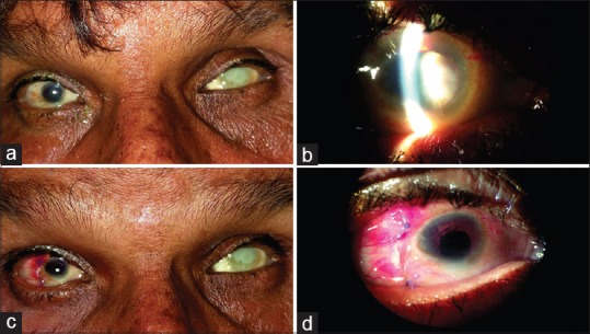
Figure 2.

(a) Microcornea of 7 mm diameter measured on slit lamp with a hard brown cataract in the right eye (b), while the left eye is phthisical. Postoperative day one picture of the same patient with a clear cornea, well-formed anterior chamber and minimal congestion (c and d)
