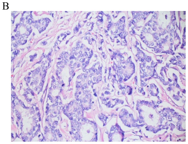
Figure 2.

Invasive ductal carcinoma obtained via lung wedge resection conducted in 2017. The tumor fills 4/5 of the photographed field and exhibits tumor replacing lung tissue, with central fibrosis surrounded by nests and tumor glands. Lung parenchyma is observed in the upper left and upper right corner (final magnification, x100).
