Figure 3.
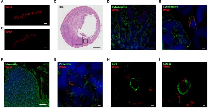
Probiotics interact with immune cells located in Peyer's Patches and villi. SIM images of WGA-AF555 (red) labeled-BA (A) and LR (B). (C) Hematoxilin and Eosin staining of rat small intestine. (D) Confocal and (E) SIM images of villi stained for cytokeratin (green) and nuclei (blue). WGA-AF555 LR (red) are located inside villi (confocal image scale bar: 50 μm). (F) Confocal and (G) SIM images of PP stained for vimentin (green) and nuclei (blue). WGA-AF555 LR (red) are located inside the PP (confocal image scale bar: 100 μm). SIM images of CD3+ cell (green) (H) and CD11c+ cell (green) (I). WGA-AF555 LR (red) are detected nearby CD3+ and CD11c+ cells. SIM image scale bar: 2 μm.

