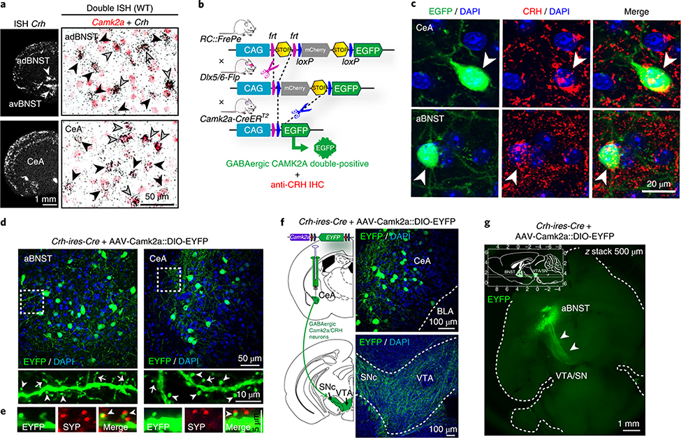
Fig. 2 |. VTA-projecting spiny GABAergic CRH neurons express Camk2a.
a, Crh mRNA expression determined by in situ hybridization (ISH, left). Double ISH (right) shows that a subset of Crh neurons in the adBNST and CeA coexpress Camk2a. Black arrowheads, Crh-positive; gray arrowheads, Camk2a and Crh double positive. Quantifications in Supplementary Fig. 3. b, Schematic representation of dual fate mapping strategy. IHC, immunohistochemistry. c, Representative sections of RC::FrePe;Dlx5/6-Flp;Camk2a-CreERT2 mice (dual-recombinase-responsive fluorescent reporter mice expressing Dlx5/6-Flpe and Camk2a-CreERT2) and subsequent CRH immunostaining (red) show triple-positive GABAergic CAMK2A CRH neurons (arrowheads) in the CeA and aBNST. d,e, CAMK2A CRH neurons carry thin (arrows) and mushroom-like (arrowheads) spines (d), which receive presynaptic input determined by synaptophysin (SYP, red) immunostaining (e). f, CeA CAMK2A CRH neurons (top) and VTA-innervating fibers (bottom). g, Whole brain CLARITY: horizontal z-stack image shows GABAergic, CAMK2A- and CRH-positive aBNST-VTA projections (arrowheads); see Supplementary Video 1. Inset shows stack range, delimited by horizontal lines; axes in millimeters, with bregma at 0 mm on the top and right axes and interaural line at 0 mm at the bottom and left axes. All experiments were independently replicated three times. Anterior dorsal BNST, adBNST; anterior ventral BNST, avBNST; basolateral amygdala, BLA.

